EQUIPMENT OF THE CENTER FOR BIOENGINERING
1. Pulsatile pump with the following capabilities:
- regulating the frequence of number of cycles per minute (50-120 cycles per minute);
- regulating inlet pressure (0-0.2 bars);
- regulating volumetric flow rate (0-1.5 litres/min).
Regulation of the above mentioned parameters is performed using the controller PIC 16F84.
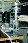
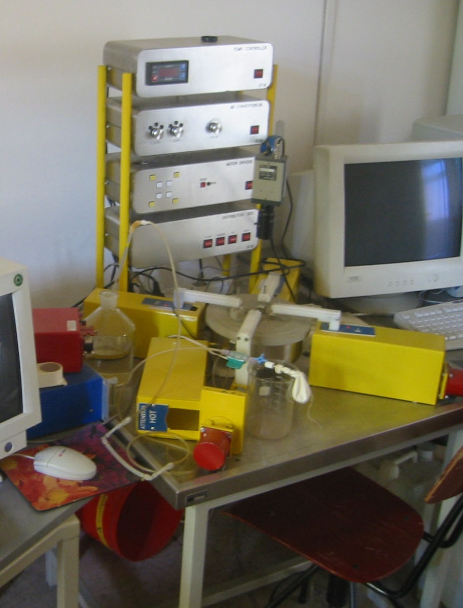
2. Experimental table for physiological testing of pulsatile flow through artificial blood vessels.
Simplified scheme of the experimental table settings is given in the figure below. Due to the cyclic repetition of the fluid flow it is necessary to form a closed-circuit flow of the process itself. In that way we enable a regularity of periodic changes and reduction of error when measuring physical quantities.
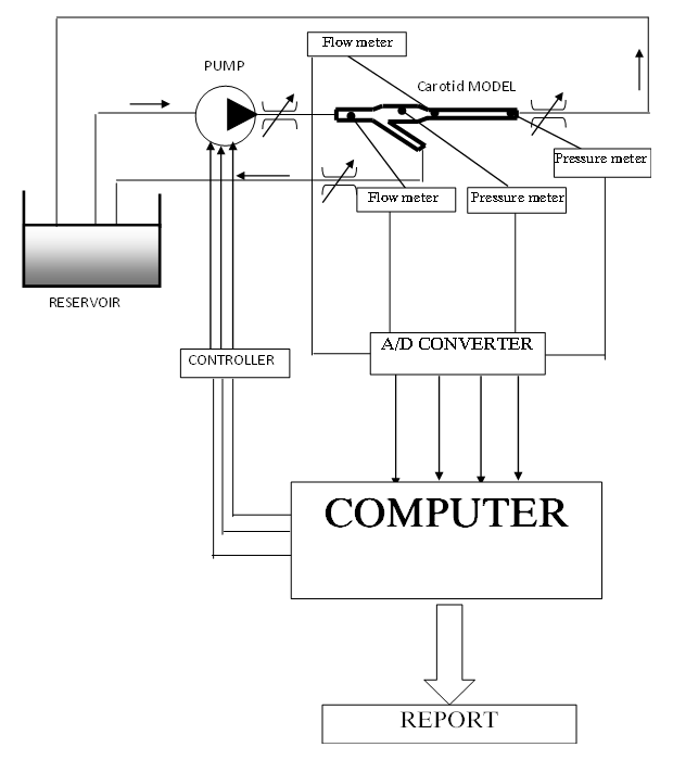
It can be seen from the picture that the pulsatile pump draws fluid from the reservoir while the given input flow in the carotid model is initiated using the computer and the controller. At the outlet of the pump it is possible to additionally regulate the flow using an orifice plate. Also, with orifice plates at the outlet branches from the external and internal carotide one can regulate the resistance force that simulate the real circulatory system (blood circulation in the face and brain). The whole system is closed in the way that the outlet of the carotidas is directed towards the reservoir where the fliud, which is used in the experiment, is placed. On the carotid model itself, at the cross section, mean velocity and mean pressure are measured. The information on quantities measured is transmitted via the A/D converter to the computer that has a major role of managing the whole system. Output values are shown on the computer display and the printing machine.
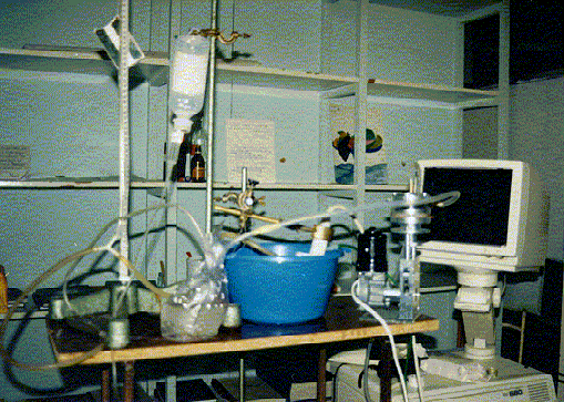
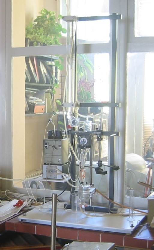
View of experimental table and connection to an ultrasound device in hospital
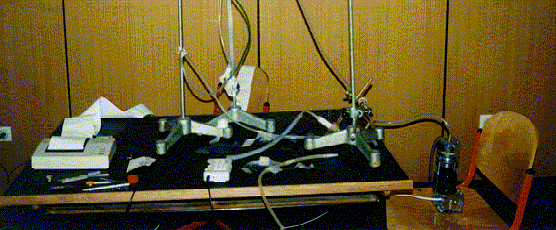
View of experimental table
3. Biaxial Stretch system for testing mechanical characteristics of tissues:
1. Mechanical part of the system consists of:
- four-axial mechanism with step motors tightening the samples,
- tub with a heater,
- table.
2. Electronic part of the system consists of:
- four controllers for controlling the step motors in the four-axial mechnaism with manual and computer control of the motor functioning,
- two bridges for measurement with amplifiers for signal processing of standard force transducer up to 50 N,
- measurment circle for the acquisition of the solution temperature,
- set for electrostimulation,
- thermoregulators regulating the tub temperature,
- block for power supply,
- CCD camera.
3. System program support enables the control of the four-axial mechanism as well as gathering the data from the amplifier of the force transducer. The program manages frequency and duration of impulses for electrostimulation. Recording is also performed throgh the program with the use of CCD camera.
4. Measuring range of force measurement from 0,01 to 50 N is satisfied on the basis of characteristics of the built-in force transducer.
5. The velocity of movement is given by the program and has the required range.
6. Frequency and impulse duration are given by the program and have the required range. Impulse amplitude is manually set but it can be monitored using the program.
4. Isolated heart system (Langendorf), EXPERIMETRIA LTD consisting of the following parts of equipment:
- two blood pressure meters,
- two blood pressure conerters,
- one temperature gauge,
- one temperature sensor,
- one extracelular signal amplifier (EKG/heart pulse),
- one perfusion device for isolated heart (with one big test tube), consisting of the following components: basic plate, supporter, big test tube, pump for filling the perfusion test tube, stimulator of the heart ventricle electrode, gas pressure regulator, silicone tubes, cables and ports.
- dispenser 0,4-2 ml
- four cannulas:
| outer diameter | inner diameter |
| 1,5 mm | 0,7 mm |
| 3 mm | 1,5 mm |
| 4,5 mm | 3 mm |
| 6 mm | 4 mm |
5. Hand-held Doppler BIDOP 3 presented in the picture below.
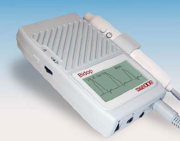
The Doppler also involves 8 MHz Sonda. Doppler has the following features:
- bidirectional measuring of systolic, diastolic and mean flow
- measuring of heart impulses 30-300 beats/min
- 30 memories
- range of frequencies: 8-20 Hz to 5 kHz
- screen resolution 128x64
- speakers volume 200 mW
6. ETH 256C – two-channel /ECG/EMG/EEG amplifier
Amplifier specification:
- number of channels: 2
- operational modes: bridge, bipotential ECG, EMG, EEG x1, x5, x10, x100, x500, x1k, x5k
- amplification x1, x5, x10, x100, x500, x1k, x5k
- low pass filters: 4-pole active filters of 5, 50, 150, 2k, and 10 khz
- high pass filters: 4-pole active filters of DC, 0.03, 0.3, and 3 Hz
- input impedance: 10 Hz
- output impedance: 100 Hz
- CMR 100 db @ 60 Hz
7. 10 PC computers, Pentium I3
8. Dynatek Dalta SVP Intravascular Stent/Graft Testing System
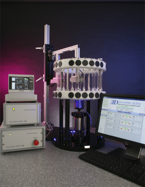
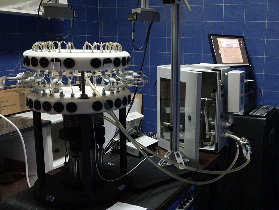
Large vascular prosthesis tester, designed to create accelerated cyclic pulsatile loading on synthetic or biological vessels. They can test up to 24 samples – radial strain testing – physiologically tuned test method (method A*). Large vascular prosthesisoperate by setting the baseline pressure, simulating the diastolic pressure, then pulsing the samples to simulate the systolic pressure using a precision bellows drive system. This pulsing, when performed under recommended conditions, closely recreates the movement that occursin vivo.The large vascular prosthesis is equipped with an external stainless steel water bath tank for sample submersion, which maintains samples at a given temperature and can create a saline environment for devices that are attached externally to your mock vessels. Due to the interchangeable manifold design, a wide variety of sizes and shapes can be accommodated on the large vascular prosthesis.This tester is designed to test samples from 6mm ID to any custom shape desired. The number of samples is determined by the shape and volume needed to achieve the target deflection.The DC7000, a microprocessor based controller allows for enhanced levels of experimental control and data capture. The DC7000 controls the speed, pressure and temperature, while recording the cycle count and allowing for graphing of the pressure signal by use of highly precise pressure transducers.
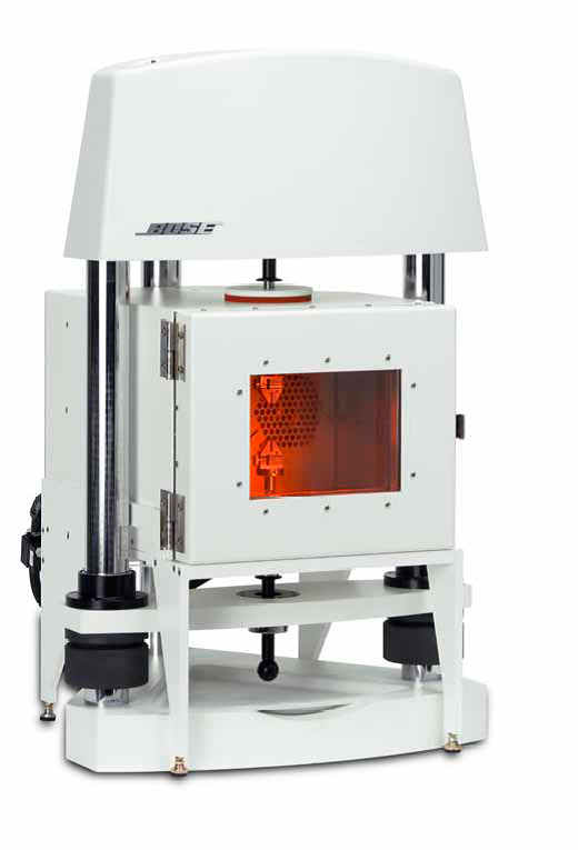
9. Bose/Electroforce/EnduraTec 9120-8 Intravascular Stent/Graft Testing System
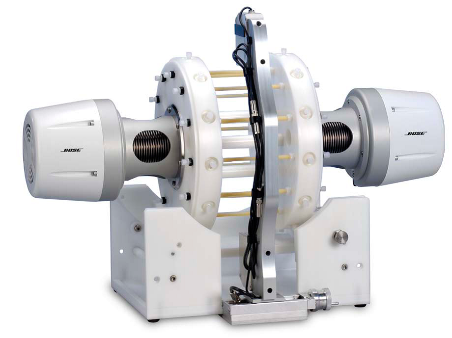
Life cycle test and radial fatigue test – a test method based on radial bending (method B*). This electrical device for stent/graft tesing verify the fatigue life of intravascular prostheses such as stents, grafts, occluders and shunts under simulated physiological displacements.The instruments are designed to provide automated control for long-term, 400-600 million cycle tests. The dual pulsatile head provides a uniform pulsatile distention at accelerated test frequencies. The system is capable of achieving a diametric distention of an artificial vessel that is equivalent to a test-to-success requirement, or, the system may be used to achieve greater distention than in vivo conditions for a fatigue-to-fracture test.This allows stent developers to calculate the fatigue life of a device design under a variety of programmed loading conditions.Options: "Pulse-on-a-Bend" for 9120-8 SGT, system for pressure and flow control with external heating system for adjusting mean pressure and constant flow through. The Bose directmeasurementsystemuses a laser/optical measurement system that is capable of providing closed loop control of pulsatile distention, or it may be used to provide real-time distention measurements on artificial vessels with a stent.
10. Bose/Electroforce/EnduraTec 3200 Multiple Sample Fatigue System
Life cycle testing and fatigue testing for other types of loads for different stents. ElectroForce® 3200 test instruments feature a 225 N (450 N optional) maximum force. With the versatility of static to 300 Hz frequency response, the configuration is adaptable to a variety of biomedical research and engineered materials test applications, including torsion testing, plastic deformation under dynamic loading and special environments (hot/cold chambers).Typical test application includes: biomaterilas, medical devices, elastomers, permeable live-tissues, microelectronics, fibres, small components, food and tissues (rheology). Highlyversatileandrobust, thesystemoperatesawiderangeofsimpleandadvancedmaterialtesting, including: tightening/pressure, axialfatiguetest, axial/torsionalfatiguetest, frictionfatigue, determination of viscoelastic properties, product endurance testing. Its flexible hardware and software platform allows for the use of multiple axes, sensors and test chambers while designing a test application.
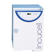
11. CO2 Incubator
Capacity: minimum 150-190 litres
- Natural air flow
- Inner glass door
- Temperature control range: from +5 °C above environment temperature to + 60 °C
- Uncontrolled relative humidity: up to max. 95 % RH at 37 °C
- Microprocessor control
- CO2 concentration: from 0.2 to 10 %
- CO2 measuring with infrared sensor and automatic calibration cycle
- Inner: 632 x 440 x 686 mm (WxLxH)±10%
- Outer: 765 x 734 x 862 mm (WxLxH)±10%
12. Inverted trinocular microscope
Optical system: infinitive optical correction system, field diameter 22 mm
Binocular or trinocular head, ocular inclination 30°, 360° rotation
Nosepiece: revolving, suitable for five objectives
Oculars: EWF
(extra wide field) oculars EW 10X /22 mm
Objective lenses:
LWD Infinity Corrected Plan Achromat Objective 4X/0,1WD 2.6 mm
LWD Infinity Corrected Plan Achromat Objective 40X/0,8WD 18 mm
Infinity Corrected Plan Achromat Phase Objective 10X/0,25WD 10 mm
Infinity Corrected Plan Achromat Phase Objective 20X/0,4WD 5.1 mm
Condenser: ELWD condenser NA 0.3. LWD 72 mm (without condenser 150 mm)
Centering: telescopic phase rings 10x-20x, phase ring plate
Specimen plate holder: Specimen plate 160 x 250 mm, glass insert of additional holder 70 x 180 mm
Focus knobs: coaxial coarse and fine adjustment; vertical sample movement
coarse movement: 37.7 mm per rotation, fine movement: 0,2 mm oper rotation
Filter: diameter 45 mm, blue, green and neutral (milk white) glass
Light source: 12V 30W halogen lamp, adjusting the light intensity
Standard additional equipment: dustcover, immersion oil, one spare halogen lamp, user manual
13. Tissue Engineering Equipment
Development of scaffolds for artificial blood vessels. Electrospinning methods, bioreactor and design of new tissue scaffolds. Computational design of tissue engineering.
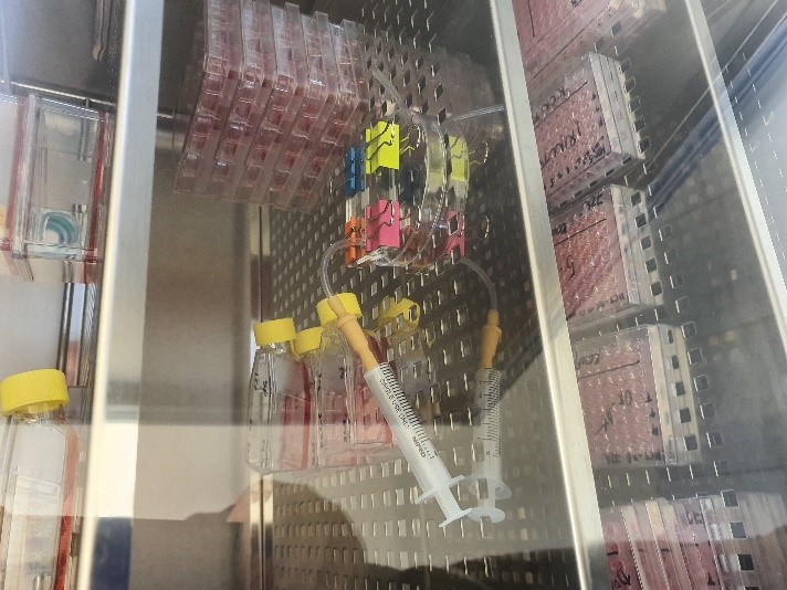
14. Cell biology Unit

The use of modern methods of cell and molecular biology in the examination of human cancer. Testing of new substances, potential preparations for cancer treatment - in vitro tests. The cell biology Unit of the Center for Bioengineering is a specially equipped system, in which human cell lines of cancer and healthy tissue are grown in optimized conditions. Methods of cytotoxicity tests, redox status of cells, gene and protein expression, micro-RNA profiling, migration, etc. they are the basis for any preclinical study, as well as monitoring the molecular mechanisms of cells in order to better understand the nature of cancer. The most current project in Cell Biology Unit is detecting of various single nucleotide polymorphisms in human COVID-19 patient samples.
15. 3D bioprint
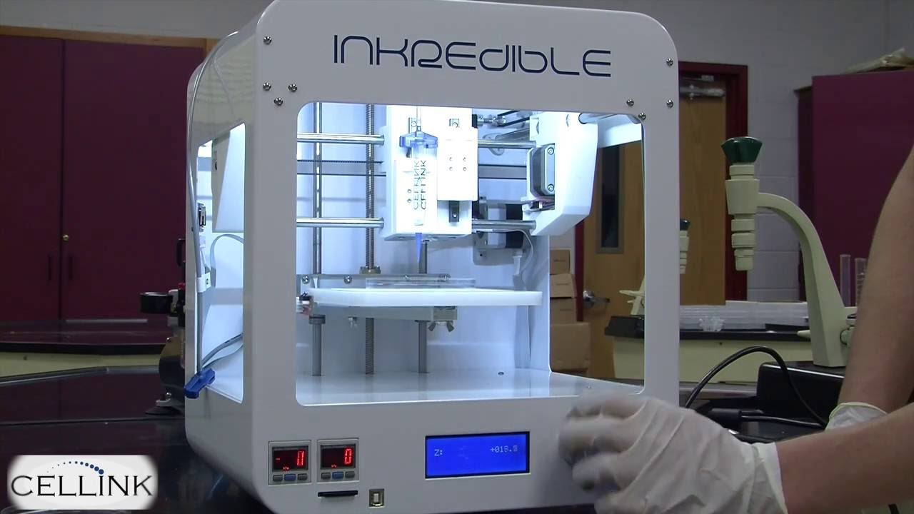
The use of in vitro techniques and animal models has significantly contributed to a better understanding of the molecular mechanisms of various human pathological conditions. However, the scope of such studies is limited. Such studies are mainly based on static testing. The Laboratory for Bioengineering creates and optimizes a real-time dynamic system with a clinically relevant assessment based on a mathematical model with and without therapy - Organ-on-a-Chip. This approach could significantly help in the early stages of clinical trials of new drugs in a complex human tissue system. One of the current projects is the creation of a Colon-on-a-Chip microfluidic system, which would mimic the affected part of the organ in the most realistic fashion possible.
16. Organ-on-a-Chip
The use of in vitro techniques and animal models has significantly contributed to a better understanding of the molecular mechanisms of various human pathological conditions. However, the scope of such studies is limited. Such studies are mainly based on static testing. The Laboratory for Bioengineering creates and optimizes a real-time dynamic system with a clinically relevant assessment based on a mathematical model with and without therapy - Organ-on-a-Chip. This approach could significantly help in the early stages of clinical trials of new drugs in a complex human tissue system. One of the current projects is the creation of a Colon-on-a-Chip microfluidic system, which would mimic the affected part of the organ in the most realistic fashion possible.
17. Electrospinning system
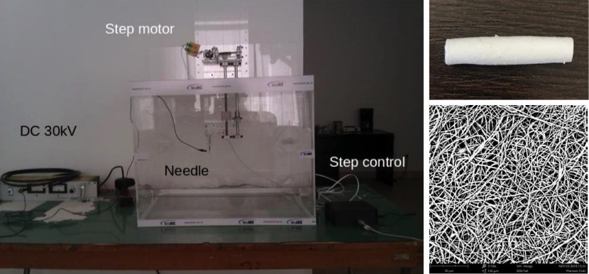
Development of scaffolds for tissue engineering. Computer-aided tissue engineering design. Scaffolds produced in this way serve as carriers on which cells are grown in special conditions, which could be transformed into mimic tissues in the bioreactor.
18. Bioelectrochemistry and biosensing
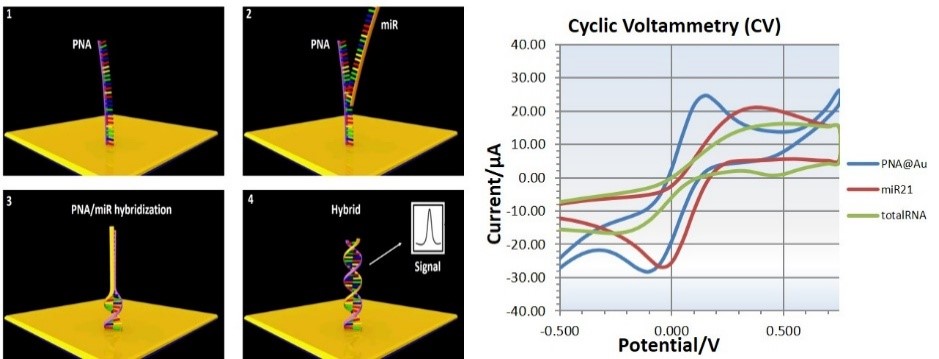
The use of electrochemistry and biosensors in the creation of devices for early detection of cancer. The goal of the development of this diagnostic device is precise and cheap detection of microRNA from blood serum or tissue without prior PCR amplification. microRNAs are considered precise markers of any pathological and healthy cell condition. Cancer cells produce different microRNAs in concentrations and ratios specific to each type of cancer. The methodology is based on hybridization of PNA with detectable microRNAs on specially created electrodes. Precise determination of microRNA content and concentration is achieved by using modern methods of electrochemistry and biosensors.
19. Stent technology development
Development of stent production technology. For this purpose, various materials of nitinol wires, their physical and electrochemical processing are examined. The characteristics of the produced stent must meet the strictest long-term patient protection regulations. Thermal and surface treatment of nitinol is extremely important, primarily due to the preparation of materials for stent production that will reduce adverse reactions in the body to a minimum.
20. PAK program
The Main Features of the Program:
PAK-G Program for graphical pre- and post- processing,
PAK-S Program for linear and geometrically and materially nonlinear structural analysis,
PAK-T Program for linear and nonlinear heat conduction,
PAK-F Program for laminar flow of incompressible fluid and heat transfer,
PAK-P Program for analysis of groundwater seepage,
PAK-CT Program for coupled problems, multiphase fluid flow with heat transfer through porous deformable medium,
PAK-B Program for biomechanics, modelling of muscles and connective tissue, blood flow through deformable arteries (solid-fluid interaction), cartilage electromechanics.
.